Skin allergies (referred to as contact dermatitis clinically) are generally a type IV hypersensitivity, also known as T cell-mediated hypersensitivity or delayed-type hypersensitivity. It takes 48-72 hours for the TH1 cells to get to the site of exposure to the allergen. The molecules that cause contact dermatitis are called haptens. Some haptens can be oxidized outside the body, called pre-haptens (examples of this kind of hapten are fragrance, dyes, and urushiol found in poison ivy). Haptens that require activation by the host metabolism are called pro-haptens. Other haptens can directly react with proteins in the body without oxidation– these are complete haptens.
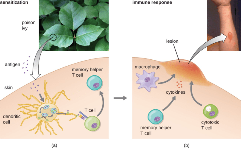
Contact dermatitis occurs when people wear or touch a hapten (like poison ivy, nickel, fragrance, etc.). A sign of contact dermatitis is induration (an inflammatory response where the skin gets thicker or harder). An example of type IV hypersensitivity is when a person’s skin touches poison ivy. The molecule urushiol, found in poison ivy, gets on the skin and goes through the epidermis into the dermis. Urushiol combines with a small self protein, forming a hapten which is recognized as non-self and is then engulfed by a dendritic cell. The dendritic cell takes the hapten to the closest lymph node. It processes the hapten proteins into peptides which it presents on its surface using an MHC class II molecule (major histocompatibility complex class type II). A naive T cell binds to MHC class II using T cell receptor (TCR) and CD4 (Co-receptor). The co-receptor is used to ensure that there will be an inflammatory immune response for that antigen. The dendritic cell releases IL-12, a cytokine that tells naive CD4+ T cell to mature into a type I Th cell –this process is called polarization. The TH1 cells releases IL-2, which helps this and other T cells activate and proliferate. Additionally, it also releases interferon-gamma (IFNγ), which helps to activate phagocytes like macrophages and create more TH1 cells. Activated macrophages release pro-inflammatory cytokines (like tumor necrosis factor, IL-1, and IL-6); this causes leakiness in endothelial vessels to allow more immune cells to enter the site of poison ivy contact, leading to swelling, edema, redness, warmth, and potentially systematic symptoms (such as fever). Activated macrophages secrete lysosomal enzymes, complement components, and reactive oxygen species to damage the tissue of the exposed area.
A subset of antigen-specific T cells become memory cells, so that the body can recognize this allergen quicker with the subsequent exposure. Antibodies against this allergen start circulating throughout the bloodstream and reach peak concentration around 10-14 days post-exposure. If you touched poison ivy again in a subsequent exposure, your body has a memory of this allergen and it is much quicker.
Definitions:
Antibodies: A blood protein that counteracts a specific antigen. Antibodies combine chemically with substances that the body recognizes as foreign.
Antigen: Any substance that causes your immune system to produce antibodies against it
Atopic Dermatitis: An itchy inflammation of the skin, also known as eczema
CD4+ T cells: They activate the cells of the innate immune system, B-lymphocytes, cytotoxic T cells, as well as nonimmune cells, and also help suppress immune reactions
Contact dermatitis: A skin rash caused by contact with a certain substance
Cytokines: small proteins that have an effect on other cells
Dendritic cells (DCs): Antigen-presenting cells (APC) in the immune system. Their main function is to process the antigen and present part of it on the cell surface to show the T cells of the immune system. They act as messengers between the innate and the adaptive immune systems
Effector T cells: T-cells that have been encountered their specific antigen and are now activated. Includes several T cell types that actively respond to a stimulus like CD4+ and CD8+
Hapten: A molecule that is incapable, alone, of causing the production of antibodies but can do so when bound to a larger antigenic molecule called a carrier.
Helper T cells: A type of white blood cell and a type of lymphocyte that stimulates killer T cells, macrophages, and B cells to make immune responses. Also called CD4-positive T lymphocyte(refer to article two for more in-depth on the difference between TH1 and TH2).
Lymph node: Lymph nodes are glands that filter out the damaged cells. Lymph nodes filter substances that travel through the lymphatic fluid, and they contain lymphocytes (white blood cells) that help the body fight infection and disease
Macrophages: Large, specialized cells that recognize, engulf, and destroy target cells
Phagocytes: a type of cell that engulfs and absorbs bacteria, particles, or other small cells
MHC class II molecule: the main function of major histocompatibility complex (MHC) class II molecules is to present processed antigens (normally found on dendritic cells)
Protein: Must be composed of one or more long chains of amino acids and are an essential part of all living organisms, especially as structural components of body tissues, enzymes, and antibodies
TCR: T cell receptor, a complex of integral membrane proteins that help in the activation of T-cells in response to an antigen
Type IV Hypersensitivity: a cell-mediated reaction that occurs in response to contact with certain allergens resulting in contact dermatitis. Certain allergens must be avoided to treat this condition
